A routine eye test can detect the earliest stages of Alzheimer’s disease in seconds, according to new research.
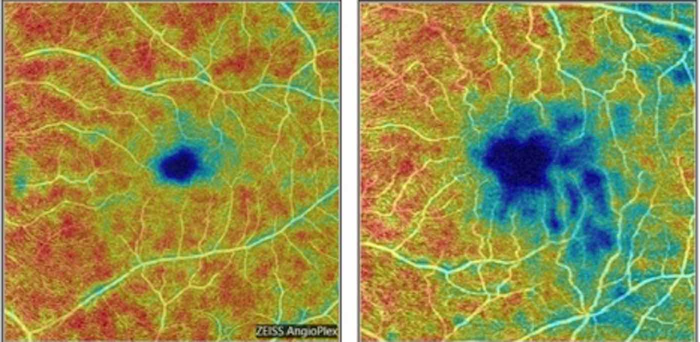
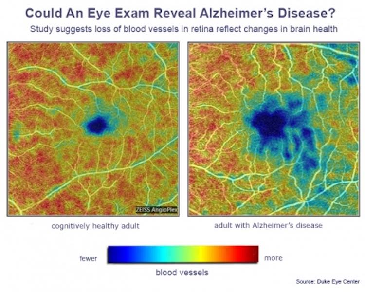
The non-invasive scan looks for changes in blood vessels in the retina. Tiny alterations in this small piece of tissue mirror those going on in the brain and are the first signs of dementia, say scientists.
The technique, which is called OCTA (optical coherence tomography angiography), could revolutionize treatment of the devastating neurological disorder because it enables early diagnosis. Physicians can now see blood vessels in the back of the eye that are smaller than the width of a human hair.
Senior author Professor Sharon Fekrat, an ophthalmologist at the Duke Eye Center in Durham, North Carolina, said: “If we can detect these blood vessel changes in the retina before any changes in cognition, that would be a game-changer.”
RELATED: ‘Eye-opening’ Dementia Breakthrough Announced Today by USC Researchers
The study published in Ophthalmology Retina compared the retinas of Alzheimer’s patients, people with mild cognitive impairment (MCI) and healthy controls.
The Alzheimer’s group had loss of small retinal blood vessels at the back of the eye. A specific layer of the retina was also thinner. Even those with MCI did not show this.
The differences in density were “statistically significant” after the researchers took into account other factors such as age and sex.
LOOK: Man Comes Up With Ingenious Way to Keep Dementia Patients Hydrated After His Grandma’s Close Call
It demonstrates how the imaging device can even distinguish between Alzheimer’s and the MCI-related preliminary forgetfulness which often leads to dementia.
The findings were based on more than 350 participants including 39 with Alzheimer’s, 72 with MCI and 254 who were cognitively healthy.
It bolsters the importance of other recent research on the eye’s connection with Alzheimer’s. It is the largest analysis of its kind to date and it adds to a growing body of evidence that OCTA is a potentially vital tool for figuring out this disease.
CHECK OUT: How Flickering Lights or Electrical Stimulation Might Banish Alzheimer’s and Parkinson’s
Because the retina is an extension of the brain which shares many of the same similarities, the tissue suggests that deterioration in one mirrors the other.
They are connected by way of the optic nerve, which Fekrat and colleagues say offers “a window into the disease process.”
The test could mean that patients may want to be given drugs decades before symptoms develop when treatments are most likely to work. Lifestyle changes, such as eating the right foods and exercising more, may also slow or even halt the condition in those identified at risk.
MORE: Alzheimer’s Gene is Neutralized in Human Brain Cells for the First Time
Current dementia medications only treat the symptoms – not the cause – meaning there is still no cure. This has led to an increasing focus on prevention.
One of the main reasons new drugs have been ineffective so far may be the fact that they are usually administered late in the disease’s development when it has already become too advanced. Early diagnosis will make possible clinical trials to administer various treatments sooner in the disease process, which may lead to better results.
Fekrat and colleagues said diagnosing Alzheimer’s is a challenge. Some techniques can detect signs of it, but they are impractical for screening millions of people.
Brain scans are expensive and spinal taps which use needles to collect cerebrospinal fluid from the spine have risks.
Instead, the disease is often diagnosed through memory tests or observing behavioral changes, by which time it’s already too advanced.
Fekrat and lead author Professor Dilraj Grewal, who are based in the same lab, expect their work will one day have a positive impact on patients’ lives.
CHECK OUT: Two Patients Make History After Essentially Being Cured of HIV Using Stem Cell Transplant
“Early diagnosis of Alzheimer’s disease is a huge unmet need,” said Fekrat. “It’s not possible for current techniques like a brain scan or lumbar puncture to screen the number of patients with this disease.
“It’s possible these changes in blood vessel density in the retina may mirror what’s going on in the tiny blood vessels in the brain. Our work is not done.
MORE: Boy is Cured of Aggressive Cancer Thanks to Stem Cell Treatment Using Donated Umbilical Cord
Her results follow a host of recent studies using OCTA that have linked eye changes to Alzheimer’s.
The cells of the retina convert light signals coming into the eye into neural signals the brain can interpret.
An earlier study of 32,000 people by University College London using a similar technique found those with thinner retinas were more likely to have problems with memory and reasoning.
SHARE the Amazing Discovery With Friends Social Media…


Except that there are also no treatments that show evidence of slowing disease progress nor are there known ways to prevent the disorder, thereby forcing the question, “Is early diagnosis really desirable”. Some people may find it to be so. But others may find that they are unable to enjoy life again after learning about their own fate. Early diagnosis would be desirable if there were ways to slow the disease progression. Since there aren’t, early diagnosis only helps researchers get closer to understanding the progression of the disorder. And that fact needs to be included in articles like this.
I believe the article indicates how this breakthrough would be helpful in trying to figure out more about this disease. The more you know about HOW a disease progresses the more CHANCES you have of stumbling upon a cure or something that staves off the effects longer.